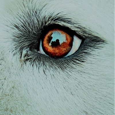
Welcome to Part 2 of this enlightening indaba with Sr. Kerryn Henson. Kerryn works as an ophthalmology nurse at an animal hospital in the beautiful Western Cape. She has so much valuable information to share that I had to divide our session into two parts. If your pet has eye issues, you’ve come to a good place!
Let’s dive right into another five challenges that can affect your pet’s eyes.
Lens luxation
Remembering that the cornea was compared to the window of the eye in Part 1, the iris can be likened to the curtains; opening and closing to let the light in-the opening being the pupil. Behind the curtain is the lens (a magnifying glass that clarifies images). The lens is suspended in place by fibres called zonules (think of an old-fashioned gong).

These ligaments assist the lens with focus by stretching and relaxing (aided by tiny muscles).
The zonules can break down so that the lens becomes completely loose (luxation). Conversely, if only a few break down it can result in the lens being suspended (subluxation). If the lens luxates, it can fall into the back of the eye or squeeze through the pupil and end up free in the space between the iris and the cornea.
The initial breakdown of the fibres can be due to a genetic defect (seen in terriers, Shar-Peis, Poodles, Beagles, Border Collies and mixes of those breeds). This leads to primary lens luxation. Additionally, some conditions like glaucoma (discussed below), inflammation, tumours and physical trauma can weaken these fibres.
When the lens detaches, it is frequently clear and therefore goes unobserved. The displaced lens can block the flow of fluids inside the globe, resulting in a build-up of pressure (glaucoma) which can be painful and causes blindness.
A luxated lens results in an odd, murky appearance of the eye and obscures a clear view of the iris. It is often accompanied by redness and pain. Treatment depends on the cause and degree of luxation and whether the lens has fallen to the front or back of the iris.
Removal of the lens is often the best solution in the case of luxation. But, because the cornea also plays a part in focusing light, pets can still see (farsighted) after this surgery. Long term medical treatment is necessary and there will always be a danger of glaucoma developing. Your vet can catch and treat any symptoms if your pet is checked regularly. Also, in the case of genetic involvement (primary luxation), there is a chance that the lens in the other eye luxates in time. Your vet can discuss treatment for that eventuality.
Glaucoma
Remember all that nutritious fluid bathing the inner eye? Well, the obstruction of ebb and flow causes a build-up of fluid inside the eye which in turn causes pressure on just about everything in there—and it’s extremely painful.
(The drainage system in the eye is a fascinating and complex system called the trabecular meshwork and drainage angle. I will spare you the details; suffice it to say that if it gets blocked, it’s bad news.)
Some breeds are genetically predisposed (primary glaucoma) and usually both eyes are affected but only one eye is impaired at a time. The list of breeds affected by glaucoma is extensive: Labradors, Spaniels, Greyhounds and Schnauzers—more often Bassets and Beagles—to name a few.
Secondary glaucoma is caused by cells and debris that block the trabecular meshwork and the drainage angle (be it a disease or physical trauma). As we have already seen, a detached lens could block the drainage angle (rapid pressure build-up), as can a tumour (slow increase in pressure). Infection and inflammation that result in blocked drainage can also cause this disorder. Bleeding inside the eye causes blood cells to block drainage too.
Signs of rapid-onset glaucoma are: discharge, rubbing and keeping the eye closed (all responses to pain). But with glaucoma, the eye bulges and becomes cloudy with a blue tinge. The whites of the eyes turn red, the pupils dilate or do not respond to light and, in the advanced stages, the animal becomes lethargic, losing appetite and ultimately losing vision. This condition requires urgent veterinary intervention. Not only is it a very painful disorder, it will damage eyesight.
Treatment aims to reduce pressure in the eye, manage the pain and promote drainage. If medical management of intraocular pressure is no longer working, the eye must be removed.
Again, getting to the vet in time could help to manage the condition and the resulting pain.
Keep an eye on those peepers for any changes!
Corneal Sequestrum
A corneal sequestrum is a part of the cornea that has died (usually in the centre). Even though the cornea does not have a blood supply, it is very similar to the skin. Dead tissue in the cornea turns necrotic and an initial vague, brown dot becomes black due to necrosis.
Although documented in horses, sequestra are most predominant in cat breeds like Persian, British Short Hair, Himalayan and Burmese. The cause is not clearly understood: continuous irritation to the cornea caused, for example, by inward turning eyelids, herpesvirus or poor ocular lubrication are all possible causes of this condition.
Apart from the dark spot, symptoms of sequestra may be blinking, squinting and light-shyness (but not always). Also, blood vessels from the edge of the eye may be seen stretching towards it. If the tissue dies back to the inner layer of the cornea, like a deep ulcer, the eyeball will leak fluids and collapse.
Lubricating ointments can manage the problem in the short term, but surgery is usually preferred because the risk of the sequestrum rupturing the cornea is a dangerous one to ignore.
The surgery to remove the sequestrum is called a keratectomy. Some surgeons will opt to cut out the spot and create a small flap of conjunctival tissue (the bright pink membrane inside the eyelids that also lines the eyeball) that is hinged towards the centre of the surgical site and stitched in place. The graft provides structural support as well as a nutritious supply of blood to the cornea. One disadvantage of the graft is that it impairs vision in the short term. But it does become thinner and clearer in a few months after surgery. Some types of grafts (not the one described) can be removed at a later stage once the cornea is healed. If your pet has had a sequestrum, it is necessary to visit your vet regularly to check for recurrence.
Top Tip:
Lubrication is luxurious! Eye drops that contain hyaluronic acid provide a longer lasting protective layer over the cornea as opposed to other lubricating drops.
Kerryn advises the regular application of preservative-free eye drops to sensitive eyes. When you take your pet out in the car or the wind, a visit to the groomer or the beach—in fact anything that can irritate sensitive eyes—pop in the drops. However, eye drops that do not contain preservatives have the potential to harbour and grow bacteria. Read the package insert and be sure to discard the bottle after the expiry date.
Cherry eye
Now here’s some interesting stuff, dogs and cats have an additional (third) eyelid. It lives in the inner corner of the eye next to the lower eyelid. It is made from conjunctival tissue stretched over a tiny T-shaped cartilage allowing the lid to sweep across the eye like a windscreen wiper.

Many types of mammals, fish, birds and reptiles employ a third eyelid to protect the eyes underwater or while hunting. Some are transparent, allowing the animal to maintain perfect vision while the lid protects, lubricates or clears the eyes. Dogs and cats however have an opaque third eyelid with a gland that contributes to lubricating the eye.
When this delicate arrangement (conjunctiva, cartilage and gland) is affected by structural changes or inflammation, the gland may become enlarged and can prolapse. This appears as a bulging in the inner corner of the eye that looks like a little cherry.
It seems that flat-faced breeds are predisposed (this is getting ridiculous!) but it also affects Beagles, Basset Hounds, Rottweilers, Saint Bernards and Spaniels. Usually, it only affects young pets (less than 2 years old).
The swelling may be uncomfortable and your pet might attempt to rub it. This causes damage to the exposed conjunctiva. It is advisable to treat cherry eye as soon as possible. Surgery is required to correct this condition.
Foreign body: grass seeds, splinters or other mechanical injuries
The last eye challenge that Kerryn talks to us about is simple mechanical damage. It is far from simple though as there are so many delicate systems that can be damaged in the eye.
As we have seen, glands, tissues, cornea and lenses all have to do a job and if they are damaged, all sorts of symptoms can occur.
Kerryn advises that any physical damage to the eye should be considered an emergency.
Specialist and expert vets have the tools to identify the type of damage and advise pet owners of their options and potential outcomes.
As we have seen, the outer layer of the cornea has a high number of nerve endings and, when damaged, the exposure of these nerve endings is very painful. It is worth bearing this in mind addressing discharge and brown stains from your pet’s eyes timeously, more serious symptoms may be avoided. Pets have a high pain threshold and only start to show signs of pain once it is serious. If you notice a change, off to the vet with you and your pet!
Thank you to Sr. Kerryn for her time and fantastic knowledge. I have learned so much from her and I believe that your pets may be better off now that you have this wonderful insight too.
Copyright Liz Roodt 2023
Co-authored and co-edited by Sr Kerryn Henson
