
Sr. Kerryn Henson shares her knowledge on eye health,
It’s a beautiful summer afternoon in the Western Cape as we share the biggest brie sandwich I’ve ever seen. Sister Kerryn Henson (previously from Gqeberha) has been working at an animal hospital in the Western Cape for a year now. Much of her time is spent assisting in the ophthalmology clinic and she has already gained such incredible insight into eyes.
Kerryn squints in the sun as if trying to organise her knowledge into a logical flow.
She begins by saying, “Sometimes owners think that tears and brown stains on the hair around the eyes (especially seen in white-haired dogs) are normal ¹. They are often symptoms indicating an underlying problem and starting early treatment will prevent more serious issues in the future.” The truth is that the sooner your pet is seen by a vet or specialist, the greater your chances are to correct disorders that could cause loss of eyesight in the future.
Corneal ulcers, ‘dry eye’, lens dislocations, displaced globes (eyeballs), deformed eyelids, glaucoma, cataracts, ‘cherry eye’ and physical damage—so much can go wrong with your pet’s eyes.
But, as Kerryn explains, it is vital to understand what qualifies any of these as an emergency.
When is an eye issue a serious issue?
Corneal Ulcers
I have heard the cornea described as the “window or windshield of the eye”; what a fantastic description for a truly magnificent little thing! A tear film (much like a waterproof barrier you might apply to your window glass) protects and nourishes the cornea. The cornea in turn has an outer layer of specialised, protective and regenerative skin-like cells, two more layers and a rich network of nerves—but notably, no blood vessels. Behind the cornea, a clear and nutrient-rich fluid keeps everything bathed and protected.
Ulcers on the cornea are commonly caused by physical trauma (from a sharp object, scratching, ingrown lashes, rubbing or chemical contact) but diseases like dry eye or diabetes can also lead to ulcers.
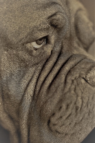
The anatomical shape of brachycephalic (flat-faced) dogs’ skulls predispose the breeds to ulcers too. These breeds suffer from many ocular diseases and are especially prone to corneal erosions. For example, many are unable to close their eyelids completely and the exposed part of the globe dries and ulcerates¹.
An untreated corneal ulcer can lead to corneal scaring and even perforation. In the case of corneal perforation prompt surgical intervention may save the eye or it may need to be removed.
Signs that your pet could have an eye ulcer are: redness, rubbing at the eye, squinting (or holding the eye closed), lots of ocular discharge and, in severely painful cases, loss of appetite and even behavioural changes. As there are no blood vessels in the cornea, you won’t see bleeding from an ulcer in the early stages.
Kerryn advises visiting the vet as soon as an ulcer is suspected. Superficial ulcers or abrasions are treatable and, once the cause has been removed, can heal quite rapidly. So, it makes sense that a timely consultation with a vet could save your pet’s eyesight.
Dry eye
Dry eye ( Keratoconjunctivitis sicca) is caused by a deficiency in the production of tears that lubricate the eye². Tears contain water, fats, proteins, oxygen, nutrition and anti-bacterial agents—all of which keep the eye moist and protected.
There are multiple reasons why glands and tear films may be dysfunctional but most commonly dry eye is caused by a faulty immune response—the immune system destroys tear-producing glands. Other causes include endocrine diseases, infectious agents, nervous system disorders and some types of medication.
Although the condition is not breed-specific, certain types of pets are predisposed to dry eye: Boston Terriers, Yorkshire Terriers, Cavalier King Charles Spaniel and English Bulldogs to name a few³.
The most prominent signs of dry eye is a thick grey to green discharge. The globe can appear crusty, dry and dull. These eyes will be red and uncomfortable and affected pets may also hold their eyes closed. The condition affects the tear film and nerve-rich cornea first, this is painful and irritating to your pet.
Dry eye may be a symptom of other disorders or diseases. Unless those issues are addressed, dry eye cannot be cured. However, treating dry eye with the correct medication —unfortunately life-long—ensures a good quality of life for your pet.
Top Tip: Sr. Kerryn says that many pet owners are afraid to remove the thick eye discharge for fear of hurting their pets. But, she maintains it’s easy to manage by using warm water and cotton wool to soften and loosen the sticky exudate.
A fine flea comb is the perfect tool to gently coax out the debris. She warns against cutting the hair—as many owners do to remove the discharge. She says, “If the eye is sensitive or painful, your pet’s first response will be to clamp it shut causing some of the surrounding hair to rub on the eye. And if the hairs are cut short the sharp ends will damage the cornea further.”
Entropion and ectropion – Eyelids turning inwards-or outwards
Many breeds suffer from entropion and ectropion. It simply relates to the amount of skin around the eyes—too much skin (as seen in Shar-Peis or flat-faced breeds) and the eyelids roll inward; heavy and drooping skin (in Bassets and Bloodhounds), and the eyelids are stretched away from the globe.
Entropion causes many symptoms (described under the section on corneal ulcers): Inflammation (redness and swelling) due to the irritation caused by the rubbing of the eyelashes on the cornea and lid spasms are potential signs of entropion as well. Treatment is surgical and the results are usually successful.
Ectropion can also be caused by immune diseases, facial nerve and muscle disorders, unresolved facial or eye injuries and even overcorrection of entropion surgery. As the delicate tissues inside the eyelid are exposed, ectropion leads to inflammation and corneal damage. Whenever the cornea is abraded, your pet experiences pain.
Treatment of these conditions is always surgical, and the prognosis is generally very good. Kerryn says that although these problems may not seem like emergencies they are considered a welfare issue. They should be addressed by a vet or specialist as soon as possible.
Cataracts
Cataracts are irreversible and usually considered to be an age-related or genetic condition. Over time, reactions occur in the lens, making it appear cloudy and white.
Cataracts can also be inherited or caused by diabetes, nutritional problems, trauma or chronic inflammation and can lead to blindness. Cataract development could lead to inflammation and in turn to glaucoma, which is so painful that it may be necessary to remove the globe.
The slow onset of cataracts is not a clinical emergency but it is worth a visit to your vet when you notice cloudiness or any other changes in the eyes. The vet nurse can provide valuable advice to pet owners about managing pets that are losing their sight.
Top Tip: Kerryn’s top tip concerning slow changes in the eye is to take a close-up picture of the problem as soon as you notice it. A chronological record will indicate the progress of changes. Photographic evidence will help the vet to understand the condition better. It should be noted that any rapid changes in the appearance of the eye should be considered an emergency.
Proptosis
This is the displacement of the eyeball outside the socket due to physical trauma (like road traffic accidents) and is a real emergency. In very few cases, surgical replacement of the globe results in restored vision. However, the damage that displaces the globe can also damage nerves, muscles and blood flow to the eye. Prognosis is usually guarded. A displaced eyeball must be treated as an emergency by seeking immediate veterinary help.
End of Part 1
Kerryn’s insights have really opened my eyes today. But there is still so much more to discuss. Join us for Part 2 of this discussion where we discuss ‘cherry eye’, lens luxation, glaucoma and a serious condition called Corneal Sequestrum in cats.
¹ Costa, J. Steinmetz, A. & Delgado. 25 January 2021. Clinical signs of brachycephalic ocular syndrome in 93 dogs. Irish Veterinary Journal. Article number:3 2021. https://irishvetjournal.biomedcentral.com/articles/10.1186/s13620-021-00183-5#citeas
² Ofri,R (DVM,PhD,DECVO). 17 March 2021. Diagnosis and treatment of dry eye in dogs. Dvm360. April 2021 Volume 54. https://www.dvm360.com/view/diagnosis-and-treatment-of-dry-eye-in-dogs
³ Hunter, T (DVM) & Ward, E(DVM). 2 December 2022. Keratoconjunctivitis Sicca (KCS) or Dry Eye in Dogs. MARS. 23 February 2023. https://vcahospitals.com/know-your-pet/keratoconjunctivitis-sicca-kcs-or-dry-eye-in-dogs
Copyright Liz Roodt 2023
Co-authored and co-edited by Sr Kerryn Henson